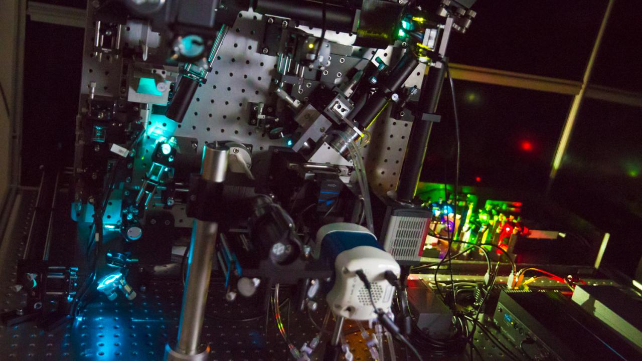
Lattice Light-Sheet Microscope Enables Discovery in Cellular Biology
The addition of a ground-breaking microscope to the College of Biological Sciences’ arsenal of research tools will transform the way UC Davis life scientists conduct research, researchers say. The lattice light-sheet microscope — one of approximately 25 of its type in the world — has the potential to revolutionize what is known about the living cell.
“Think about Galileo and his telescope,” said Michael Paddy, scientific coordinator for the Light Microscopy Imaging Facility in the Department of Molecular and Cellular Biology. “His invention changed astronomy and our understanding of our place in the world.”
The lattice light-sheet microscope has that same potential, said inventor and 2014 Nobel Prize Laureate Eric Betzig during a College of Biological Sciences Storer Lectureship in the Life Sciences presentation on campus in February. Betzig believes that with this microscope, the same level of advancements made with Galileo’s telescope will happen in cellular biology.
Powerful views
The lattice-light sheet microscope boasts many advanced capabilities over other research microscopes:
- Integrating technologies: A refined version of a light-sheet fluorescence microscope, the lattice light-sheet microscope combines techniques from Bessel beam and structured illumination microscopies.
- Increasing observing durations: The lattice light-sheet microscope allows users to view live specimens 100 to 1000 times longer than traditional spinning disc confocal techniques.
- Limiting cell damage: Most research light microscopes blast large amounts of light particles at samples that quickly damage cells in a process known as photobleaching. The lattice light-sheet microscope, on the other hand, applies an extremely thin sheet of observational light on a live cell sample, significantly decreasing cell damage.
- The process can be compared to a movie scene. Imagine only the actors speaking are illuminated, while objects in the background and foreground remain unlit. If those foreground or background objects become necessary, the director will illuminate them separately and later combine them with the images of the actors, effectively limiting the amount of light cast on the scene.
- Superior data collection: Capturing images at 50 frames per second, a sensitive camera creates detailed, continuous movies over much longer time periods, with fewer gaps in the process. This movie-like data collection process provides unprecedented three-dimensional views of changing biological processes.
Advancing scientific knowledge
“It is a transformative technology,” said Department of Molecular and Cellular Biology Chair Jodi Nunnari. “Outstanding biological questions that were previously intractable are now addressable. In short, it will enable discovery.”
Students will have significantly expanded opportunities in discovery, research application and technology access, Nunnari said, adding that the Howard Hughes Medical Institute and eight UC Davis groups collaborated to fund the $600,000 microscope, which was developed in collaboration with Intelligent Imaging Innovations of Denver, Colorado.
Together, these eight research groups raised funds for the microscope. With many departments gaining access to this new tool, UC Davis will continue its collaborative approach to advance scientific discovery.
What these discoveries might look like, however, will remain unpredictable and even unimaginable until they happen, Paddy said.
“It’s a trip into the biological unknown,” he said. “It’s a little like the (Star Trek) starship Enterprise heading out on a five-year mission. You don’t really know what’s out there until you take a look. Whether the insights will be as profound as Galileo’s, only time will tell. But it will be exciting to be on this grand adventure of taking a much closer look.”
Video: Lattice Light Sheet Microscopy
(Betzig lab/HHMI/Science magazine)
Media Resources
- This story first appeared on the UC Davis Egghead blog
